
NEW– Breakthrough –NEW
Parkinson’s: It’s an autoimmune disease!
For the first time, biomarker (antibodies) have been detected in the cerebrospinal fluid of Parkinson’s patients

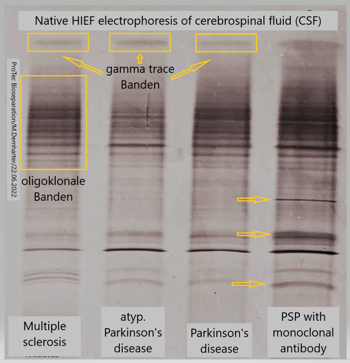
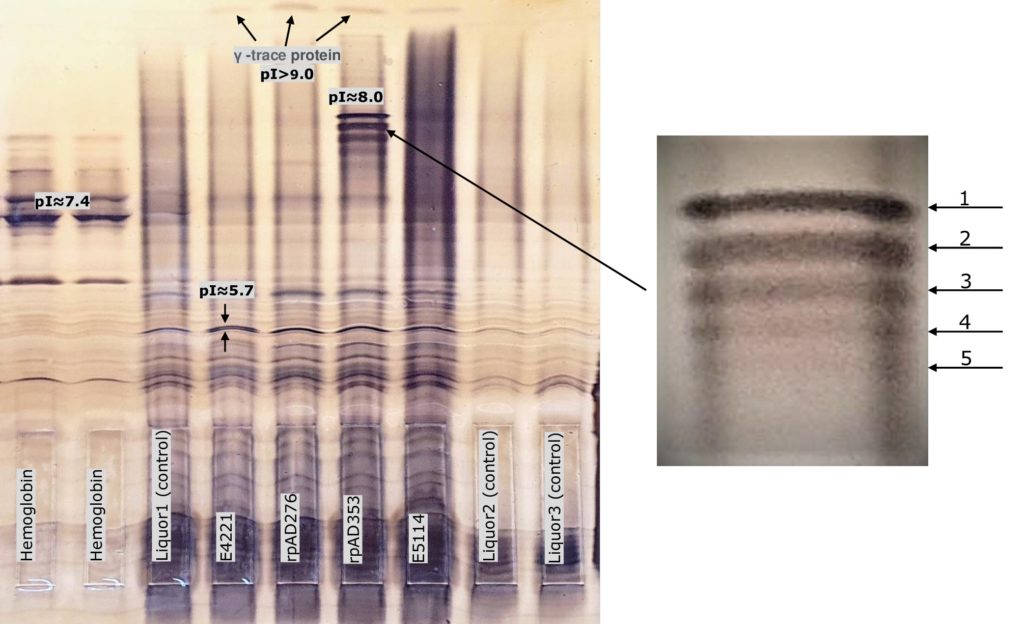
The images above is a cerebrospinal fluid (CSF) from a patient who was diagnosed with progressive supranuclear palsy (PSP)
figure left
The antibodies detected for the first time in CSF using the new HIEF electrophoresis (ProTec Bioseparation, Germany) in patients with the Parkinson’s variant PSP (Progressive Supranuclear Palsy) support the following assumptions:
For decades, researchers and physicians alike have found that inflammation drives the changes in the brains of people with Parkinson’s disease. However, it is only in recent years that they have been able to understand that this inflammation is part of the cause of the progressive nature of Parkinson’s disease and not just a consequence of the disease.
A 2017 study led by Sette and Sulzer was the first to show that alpha-synuclein can act as a beacon for certain T cells, causing them to mistakenly attack brain cells and potentially contribute to the progression of Parkinson’s. This was the first direct evidence that autoimmunity could play a role in Parkinson’s disease.
Furthermore, research by Columbia University and the La Jolla Institute for Allergy and Immunology in the United States has likewise found evidence of the role that autoimmunity plays in Parkinson’s disease. The researchers claim that two toxic fragments of the protein that appears in the brain cells of people with Parkinson’s, alpha-synuclein, can activate T cells.
According to the researchers, the autoimmunity of Parkinson’s arises when neurons cannot get rid of abnormal alpha-synuclein. This protein begins to accumulate because the process of protein recycling decreases with age and certain diseases. Since the immune system had not identified it, it was considered a pathogen to be attacked.
figure right
The very conspicuous protein double band discovered in the CSF in the anodic region of the HIEF gel (HIEF-electrophoresis) is most likely part of an oligomeric degenerate alpha-synuclein. This may be the first directly detected toxic protein, constituting evidence of PSP.
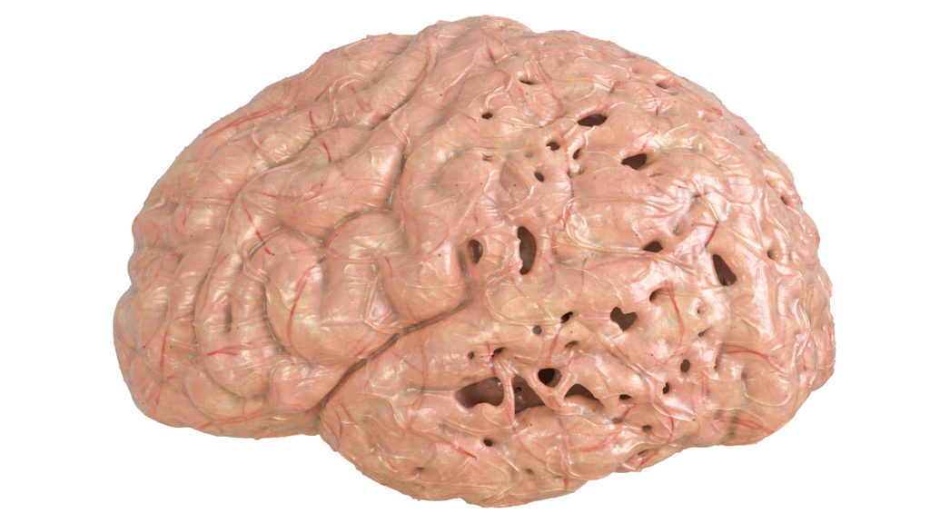
Progressive supranuclear palsy (PSP) is a rare brain disorder that causes problems with movement, walking and balance, and eye movement.
It results from damage to nerve cells in the brain that control thinking and body movement. The disorder’s long name indicates that the disease worsens (progressive) and causes weakness (palsy) by damaging certain parts of the brain above nerve cell clusters called nuclei (supranuclear) that control eye movements.
PSP is different from Parkinson’s disease—another movement disorder—although they share some symptoms. Currently, there is no effective treatment for PSP, but some symptoms can be managed with medication or other interventions.
It is well documented (Braak’s Hypothesis) and now be confirmed, that autopsied brains from patients with Parkinson’s disease (PD) show evidence of inflammation within the region of the brain thought to be responsible for PD
Logical consequence: the presence of antibodies in the CSF of PD patients.
However, such antibodies have not so far been found!
Monoclonal antibodies against Parkinson’s disease
Immune therapy approaches are widely under evaluation in most neurodegenerative proteinopathies, including Alzheimer’s and Parkinson’s disease.
There are several recognized pathways leading up to dopaminergic neuron loss in the substantia nigra and other cells in the brain as a result of age-related, genetic, environmental, and other processes.
Of these, the most prominent is the role played by the protein α-synuclein which aggregates and is the primary component of Lewy bodies, the hallmark of PD. The latest disease-modifying treatment options being investigated in PD are active and passive immunization against α-synuclein.
The main challenge is the lack of good biomarkers for target binding and the pharmacological effect of therapeutic antibodies.
The invention of the new native HIEF-electrophoresis (www.protec-biosep.de) has made it possible for the first time to detect administered monoclonal antibodies in the cerebrospinal fluid of patients with progressive supranuclear palsy (PSP).
This would allow monitoring the efficacy of new highly effective therapeutic monoclonal antibodies in the course of a medical trial and assessing the effectiveness of different antibody variants.
https://www.linkedin.com/embed/feed/update/urn:li:share:6963176595196944384
HIEF-Electrophoresis
Hydrophobic Interaction
Isoelectric Focusing
38 years of experience in the manufacturing of IEF carrier ampholytes, e.g. Sepalytes and GelStick PAG film, and sustained efforts to optimise IEF separation performance have led to the new product we call
ProPhyl Air Gel Matrix
ProPhyl Air stands for: protein loving, open structured
ProPhyl Air is a new type of gel matrix with unusual physical properties which, in combination with our self-produced carrier ampholyte (Sepalytes), represents an excellent resolution of native protein mixtures in isoelectric focusing. ProPhyl Air improves the solubility of proteins because it carries charged groups.
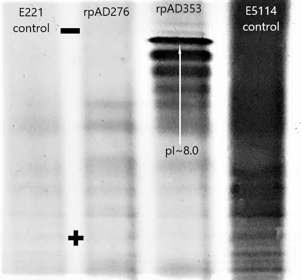
Result of Alzheimer’s samples (2nd from right) made from the BlueHorizon horizontal compartment bed-chamber to sharper bands in general and also more bands. Its use could therefore be revolutionary for those working with a wide range of proteins.
IEF Separation with ProPhyl Air
A new service for potential newcomers is a free examination of protein samples using high-resolution electrophoresis (isoelectric focusing).
Send us samples with a brief description of the material and we will send you the results within 1 week.
An advanced IEF technique for analysing native proteins
The HIEF electrophoresis technique separates molecules based on their hydrophobicity and isoelectric point. Thanks to matrices that operate under less denaturing conditions, HIEF is useful for analysing proteins while maintaining biological activity.
ProPhyl Air and its properties
The hydrophobicity of the ProPhyl Air matrix leads to an increased retention of proteins during migration compared to the common and globally used polyacrylamide. This indicates intensive interaction of the non-polar groups in the ProPhyl Air matrix with the outwardly directed polar and thus hydrophilic protein groups.
As a result of this interaction, proteins and protein mixtures that have different charge states on their outer tertiary structure are delayed in their migration to a greater or less extent, thereby producing an improved separation behavior in the ProPhyl Air matrix and also preserving the native tertiary structures of the protein.
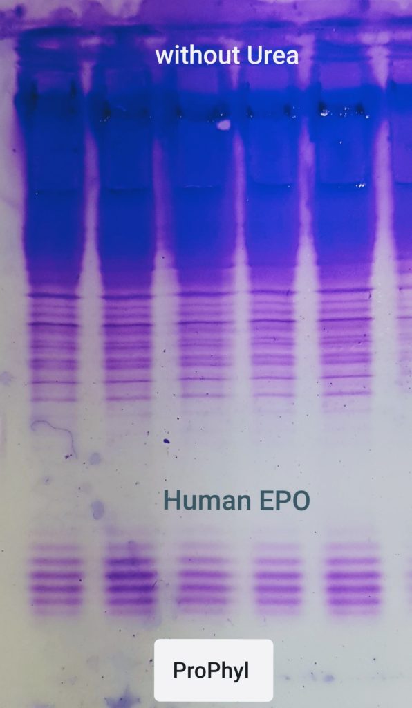
The non-polar molecules on the ProPhyl Air matrix can be referred to as „protective groups“. Based on the example of EPO and the IgG protein in CSF, our hypothesis supports/proves the fact that in the ProPhyl Air matrix the separated proteins, or the isomeric subgroups of the proteins, are shown as clear, distinct bands, in contrast to the polyacrylamide gel, in which the bands are blurred and can often only be observed as a protein cloud (denaturation).
Future Outlook
With the help of ProPhyl Airit is now possible to discover native proteins that were previously undetectable with the conventional acrylamide gel matrix used worldwide for 60 years. Examples of such proteins are biomarkers for cancer, Parkinson’s disease (PD), Alzheimer’s (AD), and EPO.ProPhyl Air also exceeds the capabilities of the IPG matrix (2D electrophoresis) in certain areas, e.g. membrane proteins, because of excellent protein solubility in the gel matrix and superior performance in the first dimension of 2D. The comparative advantages of the new ProPhyl Air gel matrix are its speed and flexibility.
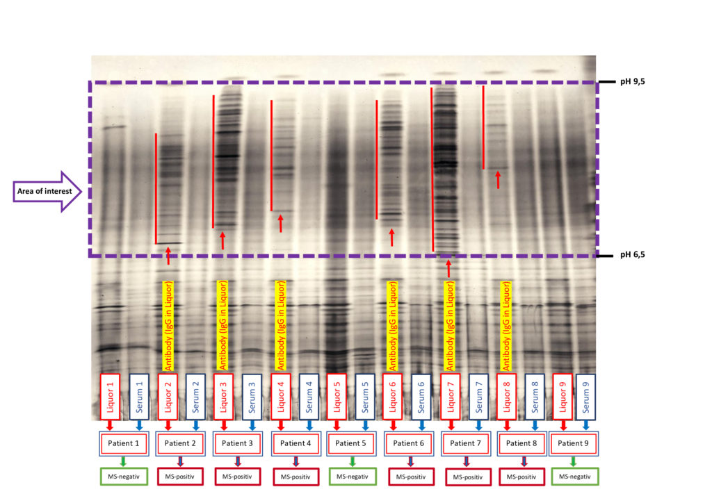
HIEF analysis of Multiple sclerosis (MS)
Improved detection of a intrathecal IgG synthesis: oligoclonal bands in multiple sclerosis
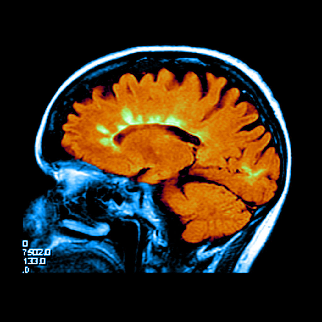
HIEF analysis of Alzheimer’s disease (AD)
HIEF analysis of Carbohydrate Deficient Transferrin (CDG)

HIEF analysis of Erythropoietin (EPO)

What is isoelectric focusing (IEF)
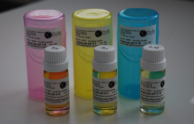
Carrier ampholytes for isoelectric focusing (IEF)
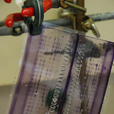
GelStick polyester sheets
– N E W –
SDS-Electrophoresis with the new gel matrix
ProPhyl Air
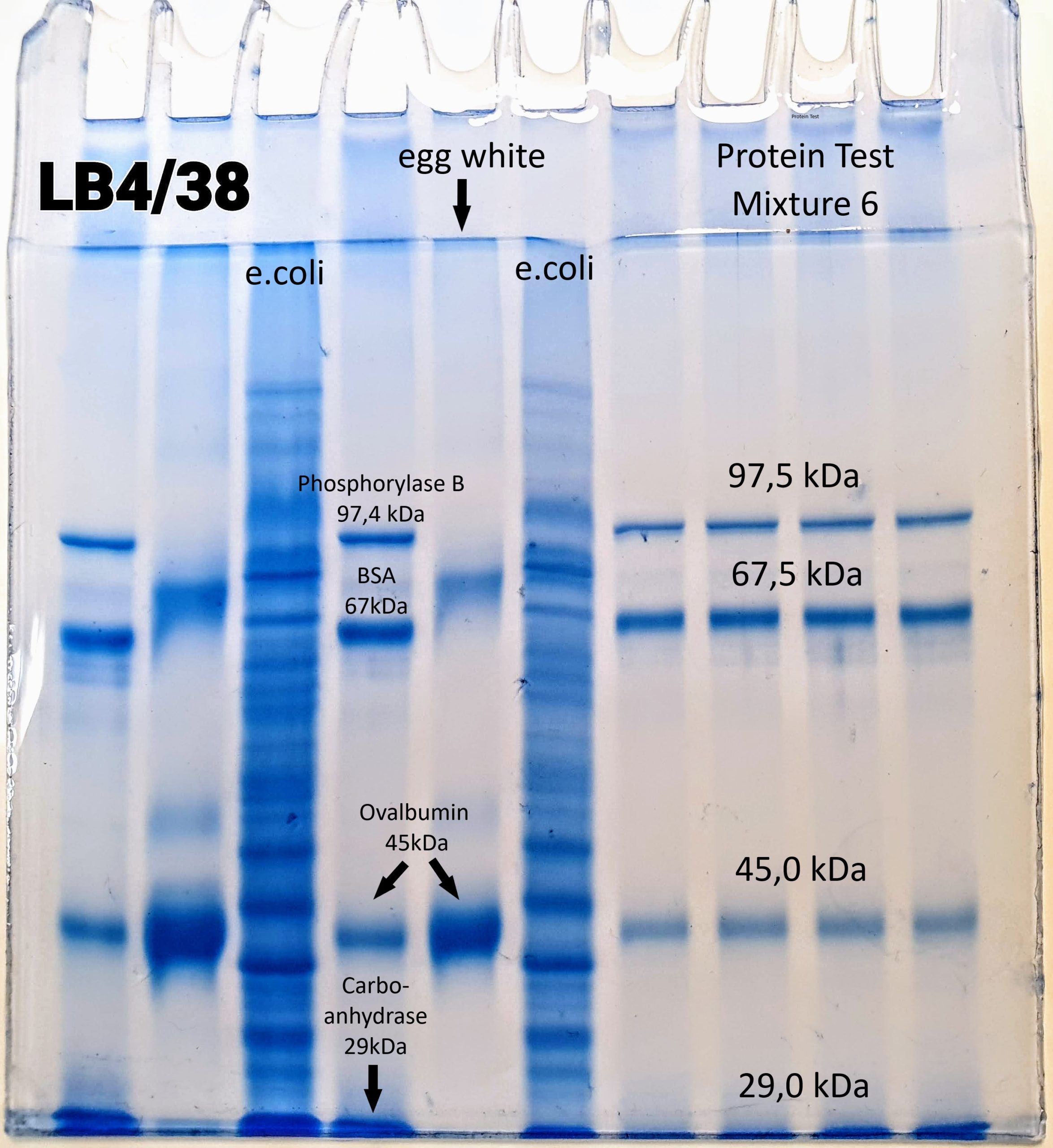
12% Tris/Glycin gel
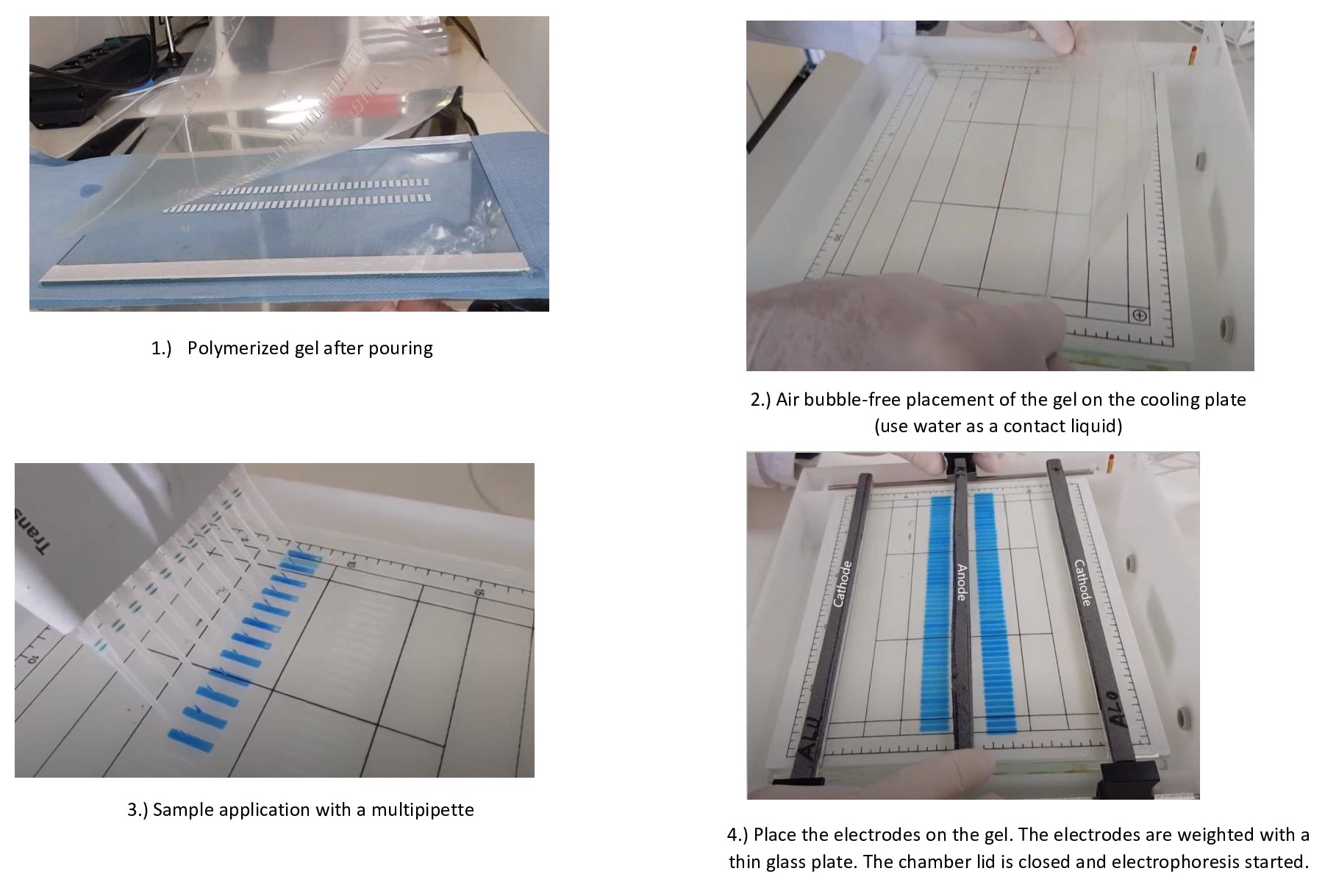
How to cast your own horizontal IEF gel
Need some advice?
No problem, just write to us!
Email: m.demharter@protec-biosep.de